
Education Portal · Latest Videos
Open Education Portal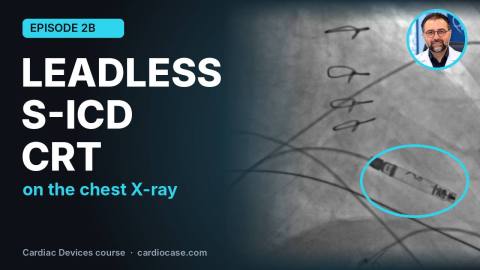
D2b_subtitled
Leadless Pacemaker, S-ICD and CRT on Chest X-ray: Special Device Systems | Cardiac Devices 2B
Question Bank · Cases
 Case #786
ICD shocks in a stable patient. Device analysis below.
Which statement is correct about the shocks?
 Case #785
A 38-year-old male with acute anterior STEMI undergoes successful primary PCI of the left anterior descending artery. Approximately two hours later, he develops a sudden wide-complex ventricular rhythm on telemetry. He is hemodynamically stable and asymptomatic. The ECG is shown.
What is the most appropriate management for this rhythm?
 Case #784
A 57-year-old woman with palpitations and manifest preexcitation and WCT in surface ECG, referred for ablation.
Surface ECG during sinus rhythm, arrhythmia, ECG during EPS before ablation, full preexcitation, arrhythmia induction and after ablation are shown, respectively.
What is the most likely diagnosis of the arrhythmia and the accessory pathway location?
Case #783
A 62-year-old man with frequent palpitations. ECG and EP tracing are shown below.
Which diagnosis is not possible?
 Case #778
This series of chest radiographs was obtained from a patient during a follow-up visit for the routine analysis of her implanted device.
Which of the following statements is incorrect based on the provided chest X-rays?

Case #782
A patient with frequent palpitations underwent EPS you can see the EP tracing below:
What is the most probable diagnosis?
Case #781
A young man with frequent PVC. Below are 3 signals from RVOT, Distal CS, and RCC.
Which signal do you think is the success signal?
Case #780
A man in his 70s presented with frequent premature ventricular complexes (PVCs) and a reduced left ventricular ejection fraction (LVEF). The morphology of his PVCs is shown below.
What is the origin of PVC?
 Case #779
A 51-year-old diabetic woman presented with a complaint of chest pain.Based on the ECG coronary angiogram performed.
Based on the ECG and Coronary angiogram,what is the next step?
Case #777
A 39-year-old man with history of DCM and reduced LVEF (25%), presented with palpitations and frequent PVC on surface ECG and Holter monitoring (PVC Burden: 40%), referred for ablation. EPS was done by 3D NAVX/Precision mapping guide. Initially, Based on surface ECG which was compatible with LV summit origin, mapping was started within CS, the best earliest signal was found in GCV (-42ms). RF applied on aforementioned area, but PVCs did not disappear. So, mapping was done via retrograde trans-aortic approach. After maintaining safe distance from LM,LCX and LAD by coronary angiography and considering correspondent point from epicardial mapping, the best earliest point was found around ILT area (-44ms). RF applied on aforementioned area and PVCs disappeared immediately.
Four fluoroscopic views and surface ECG are shown.
Which of the fluoroscopic views is in favor of catheter ablation position in the ILT (Inter Leaflet Triangle)?
Dr. Maryam Khoshnevis
MD, Cardiologist Fellowship of Echocardiography Faculty member of TUMS