Education Portal · Latest Videos
Open Education Portal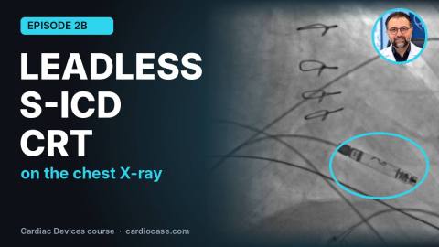
D2b_subtitled
Leadless Pacemaker, S-ICD and CRT on Chest X-ray: Special Device Systems | Cardiac Devices 2B
Question Bank · Cases
 Case #776
A 52-year-old lady with frequent palpitations, below you can find the ECG and EP tracing.
What is the most likely diagnosis?
 Case #775
A 27-year-old man known case of ACM (Arrhythmogenic Cardiomyopathy) presented with WCT in favor of VT, with history of previous unsuccessful Endocardial VT ablation, was candidated for Epicardial approach of VT ablation.
The surface ECGs (sinus rhythm & arrhythmia) are shown below.
Based on the ECG, how many major ACM criteria are met in total?
Case #774
A 55-year-old man with palpitations and WCT on surface ECG and history of successful previous VT ablation, referred for ablation. Surface ECG is shown:
Where is VT origin?
 Case #771
A 52-year-old man with a past medical history of hypertension presents with dyspnea. Myocardial perfusion imaging demonstrates reversible ischemia in the anterior wall territory. Elective coronary angiography sequence is displayed below.
Based on the most likely diagnosis, which of the following options is incorrect?
Case #772
The image below shows fluoroscopic views (LAO & RAO) of mitral annulus accessory pathway ablation via retrograde trans-aortic approach in three patients: A, B, and C
Which one is related to the Posteroseptal accessory pathway ablation?

Case #733
A 68-year-old woman with the past medical history of aortic valve replacement (AVR), mitral valve replacement (MVR), and septal myectomy (2013), presents to the emergency department. She reports a two-day history of substernal heaviness. The onset of her symptoms coincided with a interpersonal conflict involving her daughter and son-in-law.
Her prior coronary angiography, performed at the time of her valve surgery, was reported as normal.
Based on the most probable diagnosis suggested by the ECG and echocardiography findings, which of the following choices is correct?
Case #732
A young woman underwent an electrophysiology (EP) study. Interpret the tracing below.
What is WRONG about this tracing:
Case #731
A young woman with an antrospetal accessory pathway (AP) underwent an electrophysiology study (EPS) and ablation. Following the procedure, adenosine was administered to confirm the elimination of the AP pathway. The tracing below was recorded during the adenosine injection
Which statement is NOT correct about this tracing:
Case #720
A 56-year-old man with palpitations and manifest preexcitation on surface ECG, referred for ablation.
ECGs before and after full preexcitation are shown.
Where is the most likely location of the accessory pathway?
Case #719
A 58-year-old male with a history of AVR, underwent implantation of a dual-chamber pacemaker from the right side of the chest. At the time of implantation, before leads screwing (Attachment of Leads to Device header) and placing the pacemaker in the pocket , the atrial lead impedance was 650 ohms. After placing the pacemaker in the pocket, suturing, and dressing the wound, reprogramming was performed. This time, the atrial lead impedance was 1750 ohms. Chest X-rays of the patient during implantation and after implantation are available.
What is the most likely diagnosis?
Dr. Maryam Khoshnevis
MD, Cardiologist Fellowship of Echocardiography Faculty member of TUMS